Cardiogenic Shock

Cardiogenic shock is failure of the heart to pump adequately results in reducing cardiac output and compromising tissue perfusion. The treatment is focused to maintain tissue oxygenation and perfusion and improve the pumping ability of the heart.

Signs and Symptoms:

  • Hypotension
  • Urine output less than 30 mL/hour
  • Poor peripheral pulses
  • Cold, clammy skin
  • Pulmonary congestion
  • Tachycardia
  • Chest discomfort
  • Disorientation, restlessness, and confusion


Nursing Intervention:
  • Administer morphine sulfate intravenously as prescribed to decrease pulmonary congestion and relieve pain
  • Administer oxygen as prescribed
  • Intubation and mechanical ventilation if needed
  • Administer diuretic and nitrates as prescribed
  • Prepare for insertion of intraaortic balloon pump, PTCA or coronary artery bypass graft if prescribed
  • Monitor arterial blood gas levels
  • Monitor urinary output
  • Monitor distal pulses


Read more...
Heart Failure (part 2)

Emergency Interventions for Heart Failure:

  • Place the patient in high fowler position with the legs in a dependent position to reduce pulmonary congestion and relieve edema
  • Give oxygen in high concentration by mask or cannula
  • Intubation and ventilator support if needed
  • Suction fluids as needed
  • Assess and monitor level of consciousness
  • Monitor for vital signs closely
  • Monitor for hypotension
  • Monitor for dysrhythmias (using cardiac monitor)
  • Assess for edema in dependent legs
  • Insert foley catheter if needed
  • Monitor intake and output
  • Administer morphine sulfate as prescribed
  • Administer diuretics as prescribed
  • Administer digitalis as prescribed
  • Administer bronchodilator as prescribed
  • Administer additional inotropic medications as prescribed
  • Administer vasodilator as prescribed
  • Monitor weight
  • Assess for hepatomegaly and ascites
  • Monitor peripheral pulses
  • Analyze arterial blood gas
  • Monitor potassium level closely


Interventions Following Acute Episode of Heart Failure:
  • Assist the patient to identify precipitating risk factors of heart failure
  • Encourage patient to verbalize feeling
  • Instruct patient in the prescribed medications and notify physician if side effect occurs
  • Advice patient to avoid over-the-counter medication
  • Avoid large amount of caffeine
  • Diet: low sodium, low fat, and low cholesterol diet
  • Instruct patient regarding fluid restriction as prescribed
  • Avoid isometric activities that increases pressure in the heart
  • Provide patient with a list of potassium rich foods because diuretics can cause hypokalemia
  • Instruct patient to monitor daily weight and report signs of fluid retention such as edema or weight gain


Read more...

Heart Failure (part 1)

Heart failure is defined as an in ability of the heart to maintain adequate circulation because of an impaired pumping capability. The cardiac output is diminished and peripheral tissue is not perfused adequately. The congestion of the lungs and periphery will occur.

There are two kind of heart failure: acute heart failure that occurs suddenly, and chronic heart failure that develops over the time.

Type of Heart Failure:

  1. Right Ventricular and Left Ventricular Failure: Most of them begin with left ventricular failure and then progresses to both of ventricles. Left ventricular failure can progress to acute pulmonary edema that death will occur if not treated immediately.
  2. Forward and Backward Heart Failure: In forward failure, the output of the affected ventricle is inadequate and causes decreased perfusion to vital organs. In backward failure, blood backs up behind the affected failure that causes increased pressure in the atrium behind the affected ventricle.
  3. Low Output and High Output: in low output failure, the cardiac output is not enough to meet the demand. In high output failure, the heart works harder to meet the demand.
  4. Systolic Failure and Diastolic Failure: Systolic failure means the problem with contraction and the ejection of blood. Diastolic failure means the problem in relaxing of the heart and filling with blood.


Signs and Symptoms of Heart Failure


Right Ventricular Failure:

  • Pitting, dependent edema in the feet, legs, sacrum, back, and buttocks
  • Ascites form portal hypertension
  • Distended neck veins
  • Tenderness of right upper quadrant
  • Abdominal pain
  • Nausea and anorexia
  • Weight gain
  • Fatigue
  • Nocturnal diuresis

Left Ventricular Failure:
  • Cough with frothy sputum
  • Dyspnea on exertion
  • Paroxysmal nocturnal dyspnea
  • Orthopnea
  • Crackles on auscultation
  • Tachycardia
  • Pallor
  • Cyanosis
  • Fatigue
  • Confusion and disorientation
  • Signs of cerebral anorexia

Acute Pulmonary Edema:
  • Severe dyspnea and orthopnea
  • Tachycardia
  • Pallor
  • Bubbling respirations
  • Expectoration of large amount of blood tinged
  • Profuse sweating
  • Cold and clammy skin
  • Nasal flaring
  • Cyanosis
  • Use of accessory breathing muscle
  • Tahycpnea
  • Hypocapnia
  • Anxiety, apprehension and restlessness
Continued to Heart Failure (part-2)


Read more...
Myocardial Infarction

Myocardial infarction is a condition in which myocardial tissue is abruptly and severely deprived of oxygen. It can lead to necrosis of myocardial tissue if blood flow is not restored.

Physical changes in the heart do not occur until 6 hours after the infarction, the infarcted area becomes blue and swollen, and yellow and gray after 48 hours. By 8-10 days, granulation tissues form, and develop into scar over 2-3 months.


There are three location of Myocardial Infarction:
  1. Anterior or septal MI or both: due to obstruction of the left anterior descending.
  2. Posterior wall MI or lateral wall MI: due to obstruction of circumflex artery.
  3. Inferior MI: due to obstruction of the right coronary artery

Risk Factors of Myocardial Infarction:
  • Coronary artery disease
  • Atherosclerosis
  • Smoking
  • Elevated cholesterol levels
  • Hypertension
  • Obesity
  • Impaired glucose tolerance
  • Stress
  • Physical inactivity


Diagnostic Procedures for MI:

  • Total Creatine Kinase Level: rises with 3 hours and peaks within 24 hours
  • CK-MB isoenzyme: peaks within 18-24 hours after the onset of chest pain and returns to normal 48-72 hours later
  • Troponin level: rises within 3 hours and remains elevated up to 7 days
  • Myoglobin: rises within 1 hour, peaks in 4-6 hours, and returns to normal within 24-36 hours
  • LDL level: rises 24 hours after MI, peaks 48-72 hours, and normal in 7 days
  • White blood cell count: elevated WBC on 2nd day of MI
  • Electrocardiogram: ST segment elevation, T wave inversion, and abnormal Q wave


Signs and Symptoms of MI:
  • Pain: crushing substernal pain, radiate to the jaw, back and left arm. Pain occurs without any causes primarily early morning and unrelieved by rest or nitroglycerin and last 30 minutes or longer.
  • Diaphoresis
  • Nausea and vomiting
  • Dysrhythmias
  • Dyspnea
  • Feeling of fear and anxiety
  • Pallor, cyanosis, coolness of extremities


Complication of MI:
  • Heart failure
  • Dysrhythmias
  • Pulmonary edema
  • Thrombophlebitis
  • Cardiogenic shock
  • Pericarditis
  • Mitral valve insufficiency
  • Ventricular rupture
  • Postinfarction angina


Intervention in Acute Stage:
  • Assess a description of the chest pain
  • Assess vital sign
  • Assess and monitor cardiovascular status
  • Administer oxygen at 2-6 L/min by nasal cannula
  • Establish IV access
  • Administer nitroglycerin as prescribed
  • Administer morphine sulfate as prescribed (if unresponsive to nitroglycerin)
  • Obtain a 12-lead ECG
  • Administer IV nitroglycerin and antidysrhythmias as prescribed
  • Monitor thrombolytic therapy
  • Monitor for signs of bleeding
  • Administer beta blocker as prescribed
  • Monitor for cardiac dysrhythmias
  • Monitor for complication of Myocardial infarction
  • Assess and monitor distal peripheral pulses and skin temperature
  • Monitor intake and output
  • Assess respiratory rate and breath sounds
  • Monitor blood pressure closely

Intervention Following Acute Stage:
  • Bed rest for the first 24-36 hours, allow patient to stand to void or use a bedside commode
  • Provide range of motion exercises
  • Monitor the complication of MI
  • Encourage patient to verbalize feeling.


Read more...

Angina


Angina is severe chest pain resulting from myocardial ischemia. It is caused by inadequate myocardial blood and oxygen supply, obstruction of coronary blood flow, coronary artery spasm, and a condition that increases myocardial oxygen demand. There is an imbalance between oxygen supply and demand.

The treatment is focused to provide relief of an acute attack, correct the imbalance between myocardial oxygen supply and demand, and prevent the progression of the disease and further attack to reduce risk of myocardial infarction.

Types of Angina:
  1. Stable Angina: it is also called exertional angina, occurs with activities that involve exertion or emotional stress and is relieved with rest or nitroglycerin. It has stable pattern of onset, duration, severity, and relieving factors.
  2. Unstable Angina: it is also called preinfarction angina, occurs with an unpredictable degree of exertion or emotion and increases in occurrence, duration and severity over time. The pain of this angina may not be relieved with nigroglycerin.
  3. Variant Angina: it is also called Prinzmetal’s or vasospastic angina. It is results from coronary artery spasm and may occur at rest. ST segment elevation is noted on the electrocardiogram.
  4. Intractable Angina: it is a chronic and incapacitating angina that is unresponsive to interventions.
  5. Preinfarction Angina: It is associated with acute coronary insufficiency and lasts longer that 15 minutes. Preinfarction angina is a symptom of worsening cardiac ischemia.
  6. Postinfarction Angina: It occurs after an myocardial infarction, when residual ischemia may cause episodes of angina.

Sign and Symptoms of Angina:

  • Pain: slowly or quickly, mild or moderate, last lest than 4 minutes and relived by nitroglycerin or rest. Pain may radiate to the shoulders, arms, jaw, neck, and back. Pain is substernal, crushing, and squeezing.
  • Pallor
  • Dyspnea
  • Sweating
  • Dizziness and faintness
  • Hypertension
  • Palpitations and tachycardia
  • Digestive disturbances

Diagnostic Procedures for Angina:
  • Electrocardiogram: Normal during rest. ST depression or elevation and/or T wave inversion during an episode of attack.
  • Stress Test: chest pain or changes in ECG or vital signs during testing indicate ischemia
  • Cardiac Enzymes and Troponins: Normal in angina
  • Cardiac Catheterization: to provide a definitive diagnosis.

Immediate Management:
  • Assess pain
  • Provide bed rest
  • Oxygen at 3 L/min by nasal cannula
  • Administer nitroglycerin as prescribed to dilate coronary arteries and relieve pain
  • Obtain a 12-lead ECG
  • Continuous cardiac monitoring


Read more...

Coronary Artery Disease

Coronary artery disease is a condition in which one or more coronary arteries become narrowed or obstructed. It can be result from atherosclerosis and an accumulation of lipid-containing plague in arteries. This condition causes decreases perfusion of myocardial tissue and inadequate myocardial oxygen supply. The goal of treatment is to alter the atherosclerotic progression.

Coronary artery disease leads to hypertension, angina, dysrhythmias, myocardial infarct, heart failure, and death.


Coronary artery narrowing is significant if the lumen diameter of the left main artery is reduced at least 50% or if any major branch is reduced at least 75%.

Sign and Symptoms Coronary Artery Disease:
  • Chest pain
  • Palpitations
  • Dyspnea
  • Syncope
  • Excessive fatigue
  • Cough or hemoptysis
  • Possibly normal findings during asymptomatic

Diagnostic Procedure for Patient with Coronary Artery Disease:

  • Electrocardiogram: ST segment depression or T wave inversion when blood flow is reduced and ischemia occurs. ST segment elevation followed by T wave inversion when infarction occurs.
  • Cardiac Catheterization: It will show the presence of atherosclerotic lesions.
  • Blood Lipid Levels: may be elevated.

Nursing Interventions:
  • Inform patient regarding the purpose of diagnostic medical and surgical procedures
  • Assist and identify risk factors that can be modified
  • Instruct the patient regarding a low-calorie, low sodium, los cholesterol, and low fat diet with an increase in dietary fiber
  • Inform the patient that dietary changes are not temporary but maintained for life
  • Instruct the patient regarding prescribed medications
  • Instruct the patient regarding exercise, smoking reduction, and stress reduction

Surgical Procedures for Coronary Artery Disease:
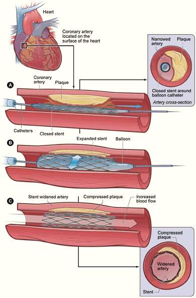
  • PTCA: to compress the plaque against the wall of the artery and dilate the vessel
  • Laser angioplasty: to vaporize the plaque
  • Vascular stent: to prevent the artery from closing and to prevent restenosis
  • Atherectomy: to remove the plaque from the artery
  • Coronary artery bypass graft: to improve blood flow to the myocardial tissue


Medications
  • Nitrates: to dilate the coronary arteries and to decrease preload and afterload
  • Cholesterol-lowering medications: to reduce the development of atherosclerotic plaques
  • Calcium channel blockers: to dilate coronary arteries and reduce vasospasm
  • Beta blocker: to reduce blood pressure


Read more...

CARDIAC TAMPONADE

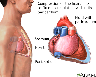
Cardiac tamponade is compression of the heart due to critically increased volume of fluid in the pericardium. The space between the parietal and visceral layers of the pericardium are filled with fluid. Acute cardiac tamponade occurs when small volume (20-50 ml) of fluid accumulate in the pericardium.

Cardiac tamponade will restrict ventricular filling and cardiac output drops.


Signs and Symptoms of Cardiac Tamponade:
  • Pulsus paradoxus
  • Jugular venous distention with clear lungs
  • Increased CVP
  • Distant, muffled heart sounds
  • Cardiac output is decreased

Intervention for Cardiac Tamponade:
  • Place patient in a critical care unit for hemodynamic monitoring
  • Administer fluid intravenously as prescribed
  • Prepare patient for chest x-ray, echocardiogram, and pericardiocentesis (to draw pericardial fluid) if prescribed


Read more...

Rheumatic Fever


Rheumatic fever is a systemic inflammatory autoimmune disease occurring after a group A beta-hemolytic streptococcal infection: pharyngitis, strep throat, scarlet fever, tonsillitis, approximately 2-6 weeks prior. Rheumatic fever can involve the heart, joints, skin, and brain and most often seen in children and young adults.

Signs and Symptoms:
  • Fever
  • Joint pain, migratory polyarthritis
  • Joint swelling; redness, or warmth
  • Abdominal pain
  • Skin rash (erythema marginatum)
  • Aschoff bodies, inflammatory hemorrhagic bullous lesions located on the myocardium usually found on autopsy
  • Sydenham’s chorea also called St.Vitus Dance—emotional instability, muscular weakness and rapid, uncoordinated jerky movements affecting primarily the face, feet, and hands
  • Epistaxis (nosebleeds)
  • Carditis
  • Subcutaneous nodules located on extensor surfaces of knees, elbows, and knuckles
  • Cardiac murmur

Treatments:
  • Anti-inflammatory medications such as aspirin or corticosteroids;
  • Antibiotic therapy (penicillin, erythromycin), includes the continuous use of low dose antibiotics to prevent recurrence; supportive therapy for other symptoms

Nursing Interventions:

  • Assess and monitor for risk factors
  • Monitor vital sign
  • Assess laboratory values for elevated erythrocyte sedimentation rate, C-reactive protein
  • Assess and monitor heart sounds and ECG
  • Administer pain medication as prescribed
  • Administer antibiotics as prescribed
  • Maintain bed rest during acute stage
  • Maintain fluid balance
  • Provide emotional support
  • Provide client and family education especially for prophylactic antibiotic therapy to reduce the risk of recurrent rheumatic fever
  • Instruct client and family in proper and prompt treatment of strep throat and scarlet fever
  • Provide client and family education


Read more...

Endocarditis

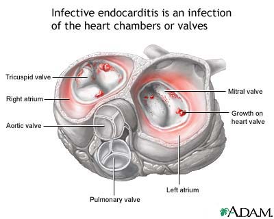
Endocarditis is the infection of the endocardial suface of the heart including the valves. Most people who develop endocarditis have underlying valvular heart disease. The disease due to rheumatic heart disease, prostatic heart valve, IV drug use, mitral valve prolapsed, and infection by bacteria (Streptococci, Enterococci, or Staphylococcus aureous). Infective endocarditis has a 20-30% mortality rate.

Endocarditis can be diagnosed by positive blood cultures and presence or valvular vegetations on echocardiogram.


Signs and Symptoms:
  • Anorexia
  • Malaise
  • Fever
  • Fatigue
  • Night sweats
  • Heart murmurs
  • Wight loss
  • Elevated ESR and WBC

Treatments:

  1. Antibiotic to eliminate all microorganism and prevent complication
  2. Surgical repair or replacement of damaged valves.

Nursing Interventions:
  • Assess and monitor vital signs and heart sound (the presence of murmurs)
  • Administer IV antibiotics as prescribed
  • Monitor for side effects of antibiotic (renal or ototoxicity)
  • Assess and monitor signs of heart failure, embolic event, or dysrhythmias
  • Provide adequate rest
  • Teach patient to avoid recurrence of infections
  • Teach patient to avoid excessive fatigue and to stop activities that result in chest pain, faintness, or dyspnea
  • Teach patient the importance of completing medication schedule and side effects of medication


Read more...

Myocarditis


Myocarditis is the inflammation of the myocardium. Generally myocarditis is due to infection from virus, bacteria, protozoa or helminthes, toxins, systemic disease, radiation, and drugs. The inflammatory process causes edema and damage to the cell of the heart that results in weakening anf the heart muscle and then decreases its contractility. Mycardtis has a 20% to 30 % mortality rate.

Signs and Symptoms of Patient with Mycarditis:
  • Anorexia
  • Malaise
  • Fatigue
  • Fever
  • Weight loss
  • Night Sweats
  • Heart murmurs
  • Elevated ESR and WBC
  • Positive blood cultures, reflecting an infectious or inflammatory

Nursing Intervention for Patient with Myocarditis:

  • Monitor vital sign and assess for presence of murmus
  • Administer antibiotics as prescribed and monitor for side effects
  • Assess and monitor for signs of heart failure
  • Assess for signs of embolic or dysrhythmias
  • Provide adequate rest periods for patient

Special Teaching for Patient with Myocarditis:
  • Teach patient how to avoid recurrence of infection: using antibiotic prophylaxis for dental or urological procedures.
  • Teach patient the side effects of each myocarditis mediction.
  • Teach patient how to modify lifestyle to avoid excessive fatigue and to stop activities that result in chest pain, faintness, or dyspnea.
  • Teach patient the importance of completing mycarditis medication schedules.


Read more...

Pericarditis


Pericarditis is the inflammation of the layers of the pericardium. It may involve the diaphragm and can be primary or secondary to other disease. Pericardium is the fibrous sac surrounding the heart.

Pericarditis may result from bacterial, viral, or fungal infections, immunologic disorders, connective tissue disease, neoplasms, renal failure, myocardial infarction, myocardial injury, radiation, or drugs.

Patient with pericarditis will show up the signs and symptoms as followed as here:

  • Chest pain that worsens when deep breathing or lying in a supine position and improves when sitting up taking shallow breaths (this is one means to discriminate from acute MI since chest pain in MI is usually not effected by change in position)
  • Dyspnea
  • Malaise
  • Fever
  • Cough
  • Elevated ESR and WBC
  • Pericardial friction rub heard most commonly on expiration
Nursing Interventions for patients with pericarditis:
  • Assess and monitor vital signs and heart sound.
  • Administer pain medication to treat malaise and other flulike symptoms as prescribed.
  • Administer NSAIDs as prescribed.
  • Give patient time for rest.
  • Assess and monitor the signs of decreases cardiac output.
  • Comfort patient: increased fluids, rest periods, and distraction technique.
  • Teach patient to adhere to medication schedules.
  • Guide patient to adopt ways to modify lifestyle especially to conserves energy and reduce fatique during acute episode of illness.
  • Teach patient how to recognize signs of recurrence: chest pain, malaise, and fever.


Read more...

Burn Wound Care: Opened Method vs. Closed Method

Opened Wound Care

The antimicrobial cream is applied every 12 hours and wound is left open to the air without any dressing.

Advantages:
The wound can easily visualized, easier mobility and joint range of motion, and simplicity in wound care.

Disadvantages:
Increase chance of hypothermia from exposure.

Closed Wound Closure

Gauze dressings are wrapped carefully from the distal to the proximal are of the extremity to ensure circulation is not compromised. The dressings are changes every 8-12 hours.

Advantages:
Using this method will decreases evaporative fluid, decreases heat loss and aids in debridement.

Disadvantages:
The disadvantages of this method are mobility limitation, prevents effective range of motion exercises, and limited of wound assessment.


Read more...

Burn Wound Care: Wound Closure

Wound closure of burn area is performed on the fifth to twenty-first day. It is depend on the extent of the burn area. Wound closure can prevent infection and loss of fluid, promotes healing and prevents contractures.

There are two types of wound closure techniques: temporary wound covering and permanent wound covering (autografting).

Temporary Wound Covering

1. Biological

Amnion:
The amniotic membranes from human placenta are used, and dressing is changes every 48 hours with amnion.

Allograft Homograft:
The donated human cadaver skin is used. Extra monitor for wound, sign of infection and sign of rejection. The rejection can occur within 24 hours.


Xenograft Heterograft
Xenograft over granulation tissue is replaced every 2 to 5 days until the wound heals naturally or until closure with autograft is complete.

Biosynthetic and Synthetic
Visual infection of wound is possible because dressing are transparent or translucent. Wound exudates and signs of infection should be monitored.

2. Autografting
Autografting is surgical removal of a thin layer of the client’s unburn skins, which then is applied to the excised burn wound that provide permanent wound coverage. This procedure is performed in the operating room under anesthesia. Bleeding following the graft should be monitored because bleeding beneath an autograft can prevent adherent. Autograft areas are immobilized 3 to 7 days following surgery to allow time to adhere and attach to the wound bed.

There are four types of skin grafts:

  • Split Thickness: Graft of half of the epidermis, applied in sheets or postage stamp-like pieces.
  • Full Thickness: Graft consisting of epidermis and dermis are used for reconstructive surgery months or years after the initial surgery.
  • Pedicle Flap: This is used for reconstructive surgery months or years after the initial inury.
  • Cultured Epithelium: The client’s unburned skin is used. Isolation of keratinocytes and culturing of epithelial cells in a laboratory and then are attached to the burn wound.

CARE TO THE GRAFT SITE:
  1. Immobilize and elevate graft site and keep from pressure.
  2. Avoid weight bearing
  3. Remove exudates (if any) using roll a cotton-tipped applicator over the graft to prevent infections.
  4. Monitor for foul-smelling drainage, increased temperature, fluid accumulation, hematoma, and increased WBC.
  5. Instruct the client to lubricate healing skin with cocoa butter as prescribed, avoid using fabric softeners and harsh detergents in the laundry, protect the affected area from sunlight, and use splints and support garment as prescribed.

CARE FOR THE DONOR SITE:
  • The methods of care for the donor site may varies depending on physician’s preference.
  • A moist gauze dressing is applied at the time of the surgery to maintain pressure and stop any oozing.
  • Keep the donor site clean, dry, and free from pressure.
  • Apply lubricating lotions to soften the area and reduce the itching.
  • Prevent the client from scratching the donor site.


Read more...

Burn Wound Care: Hydrotherapy and Debridement

HYDROTHERAPY
Burn wound area are cleansed by immersion, showering or spraying and no more than 30 minutes to prevent increased sodium loss through the burn wound, heat loss, pain, and stress. Client should be premedicated before procedure. Bleeding should be minimize and maintain body temperature during procedure.

Hydrotherapy generally is not use for patients who are unstable or those with ne skin grafts. So if hydrotherapy is not use, burn wound areas are washed and rinsed in bed before the application of antimicrobial agents.


DEBRIDEMENT
Burn Debridement means the excision of devitalized tissue and foreign matter from burn wound area. It can prevent bacterial proliferation and promote wound healing. Debridement may be mechanical, enzymatic, or surgical.

Mechanical:
This mechanical technique uses scissor and forceps to lift and trim away loose eschar. It is a painful procedure and may be bleeding and requires a moist environment to be effective. Dressings are applied directly to the burn wound area: wet-to-dry or wet-to-wet dressing technique.

Enzymatic:
Enzymatic technique means using prepared proteolytic and fibrinolytic topical enzymes that digest necrotic tissue, which facilitates eschar removal.

Surgical:
The eschar area is excised and covered. It may be tangential (shaving of thin layers of eschar until viable tissue is reached) or fascial (used for deep burns and removal of burn tissue and underlying fat down to the fascia).


Read more...

Burn Injury Management - Phase

1. EMERGENT PHASE
The emergent phase begins at the time of injury and ends with the restoration of capillary permeability (fluid resuscitation): 48-72 hrs following the injury. Key point is to prevent hypovolemic shock and preserve vital organ functioning. It includes prehospital and emergency room care.

Pre-hospital Care

  • Remove source of the burn
  • Assess airway, breathing, and circulation,
  • Conserve body heat
  • Cover burns with sterile or clean cloths
  • Remove jewelry and clothing
  • Intravenous fluid (if needed)
  • Quick transport

Emergency Room Care
It is a continuation of care administered at the scene and implemented as the hospital policy or standard procedures.


For Major burns:
For Minor Burns:

2. RESUSCITATIVE PHASE

It begins with the initiation of fluids and ends when capillary integrity returns to near normal. Keys point is to prevent shock by maintaining adequate circulation blood volume and vital organ perfusion. And the successful fluid resuscitation is evaluated by stable vital signs, adequate urine output (30-50 mL/hr) palpable peripheral pulses, and a clear sensorium.

Fluid replacement is calculated from the time of injury not from the time of arrival at the hospital. The amount of fluid given is based on the body weight and extent of the injury.

Common Fluid Resuscitation Formulas (24 hours after burn injury)

Parkland (Baxter) Formula:
  • 4 mL/kg per percent TBSA burned.
  • Half is given in first 8 hours
  • One-quarter each next 8 ours
  • Solution: Lactated Ringer’s

Modified Brooke Formula:
  • 2.0 mL/kg per percent TBSA burned
  • Half is given in first 8 hours
  • Half in next 16 hours
  • Solution: Lactated Ringer’s

3. ACUTE PHASE

It begins when the client is hemodynamically stable, capillary permeability is restored, and diuresis has begun, usually 48-72 hours after the time of injury. Key point is on infection control, wound care, wound closure, nutritional support, pain management, and physical therapy.


Read more...

Inhalation Injuries

There are four types of inhalation injury that most occurred: Smoke inhalation injury, carbon monoxide poisoning, smoke poisoning, and direct thermal heat injury.

Smoke Inhalation Injury
Smoke inhalation injury is occurred when the person is trapped in an enclosed, hot, smoke filled space. Patient with smoke inhalation might be have facial burns, erythema, swelling of oropharynx and nasopharynx, flaring nostrils, hoarse voice, stridor, wheezing, dyspnea, sooty (carbonaceous sputum), cough and tachycardia.


Carbon Monoxide Poisoning
Carbone monoxide is a colorless, practically odorless, and poisonous gas formed by the incomplete combustion of carbon; its toxic action is due to its strong affinity for hemoglobin, myoglobin, and the cytochromes, reducing oxygen transport and blocking oxygen utilization.
Oxygen molecules are displaced and carbon monoxide reversibly binds to hemoglobin to form carboxyhemoglobin that tissue will be hypoxia.

The signs and symptom of carbon monoxide poisoning are depended on the level of carbon monoxide in blood. It will be from impaired visual activity, headache, nausea, vomiting, dizziness, syncope, tachypnea, tachycardia, until coma or death.

Smoke Poisoning
Smoke poisoning is injury result when the victim inhales by-products of combustion. The localized inflammatory reaction occurs that causing a decrease in bronchial ciliary action and decrease in surfactant. The victim will have mucosal edema and wheezing.

Direct Thermal Heat Injury
Direct thermal heat injury can occur to the lower or upper airways by the inhalation of steam or explosive gases. Mucosal edema can lead to upper airway obstruction especially during the first 24 hours.


Read more...

Burn Injury Management

There are four phases of Burn Injury Management: Emergent phase, resuscitative phase, acute phase, and rehabilitative phase.

Emergent Phase

  • It includes pre-hospital care and emergency room care and begins at the time of injury till the restoration of capillary permeability.
  • Usually 48-72 hours following burn injury.
  • The management is to prevent hypovolemic shock and preserve vital organ functioning.

Resuscitative Phase
  • The resuscitative phase begins with the initiation of fluids until capillary integrity returns to normal level.
  • Administration of fluid is based on the body weight and extent of injury, and the formulas are calculated from the time of injury and not from the time of arrival at the hospital.
  • Management of this phase is to prevent shock by maintaining adequate circulating blood volume.


Acute Phase
  • This phase begins when the victim is hemodynamically stable, capillary permeability is restored, and diuresis has begun and continues until wound closure is achieved.
  • Usually 48-72 after the time of injury.
  • Management of this phase focus on infection control, wound care, wound closure, nutritional support, pain management, and physical therapy.

Rehabilitation Phase
  • This is final phase of burn management.
  • It focuses that the patient can gain independence and achieve maximal function.


Read more...

Burn Injuries (Part 3)

Rule of Nines

This method is used in calculating body surface area involved in burns. Here are Rule of Nines (Adult):
  • Head and nect: 9%
  • Anterior trunk: 18%
  • Posterior trunk: 18 %
  • Arm (9% each): 18%
  • Legs (9% each): 18%
  • Perineum: 1%


We can estimate the body surface area on an adult that has been burned by using multiples of 9.
For example, if both legs (18% x 2 = 36%), anterior trunk (18%) and both of arm (18%) were burned, this would involve 72% of the body.


Read more...

Burn Injuries (part 2)

Types of Burn Injuries

There are four major types of burns: thermal burns, chemical burns, electrical burns, and radiation burns.

Thermal Burns
Thermal burns are caused by exposure to flames, hot liquids, steam or any hot objects.

Chemical Burns
Chemical Burns are caused by contact with strong acids, alkalis, or organic compounds.

Electrical Burns
Electrical burns are caused by an exogenous electric shock that passes through the body. Muscle and tissue damage will be occurred particularly in high-voltage electrical injuries. The voltage, type of current, contact site, and duration of contact are important to identify the quick action. Just to be remembered that alternating current is more dangerous than direct current because it associated with cardiopulmonary arrest, ventricular fibrillation, titanic muscle contraction and bone fracture.


Radiation Burns
Radiation burns are caused by exposure to ultraviolet light (sun exposure), x-rays, or a radioactive source. Sun exposure is the most common burn, specifically two wavelength of light UVA and UVB that may more dangerous.


Read more...

Burn Injuries (part 1)

A burn injury is a type of injury that may be caused by heat, cold, electricity, chemicals, light, radiation, or friction.

Burn Size:

  1. Small burns: Localized burn to the injury area.
  2. Large burns: Consist of 25% or more of the total body surface area, and the response is systemic means all of the major systems of the body is affected.

Burn Classification by Depth:

1. Superficial-thickness Burn
It is similar to first-degree burn. There is mild to severe erythema but no blisters. Burn is painfull, and is ceased by cooling. It heals in 3 -7 days.


2. Partial-thickness Superficial Burn
It is similar to second-degree burn. Large blisters cover an extensive area and edema is present. Mottled red base and broken epidermis with a wet, shiny, and weeping surface. Burn is painful and sensitive to cold air. Superficial partial thickness burn heals in 2-3 weeks while the deep partial thickness heals in 3-6 weeks. If the healing process is prolonged, the grafts may be used.

3. Full-thickness Burn
It is similar to third-degree burn. A deep red, black, white, yellow or brown area are present with edema. Burn causes tissue disruption with fat exposed and spontaneous healing will not occur. There is little or no pain of burn area. It requires removal of eschar and split or full thickness skin grafting. The healing will be weeks to months.

4. Deep Full-thickness Burn
It is similar to fourth-degree burn. It involves injury to the muscle and bone. The injured area appears black, no edema, no pain and no blisters. The eschar is hard and inelastic, and healing take weeks to months. Graft are required.


Read more...

Decubitus Ulcer

Decubitus ulcer is an impairment of skin integrity when staying in one position for too long without shifting weight. It is also called as bedsore or pressure ulcer. This case often happens when using wheelchair and bedridden. The common places for decubitus ulcer are over bony prominences such as the elbow, heels, hips, ankles, shoulders, back, and the back of the head.

The constant pressure against the skin can reduce the blood supply to that area and will affect tissue dies. It will start as reddened skin and will progressively worse to form a blister, open sore, and a crater. The important thing is to prevent of skin breakdown, particularly in caring or the bedridden or immobile patient.

There are factors that increase the risk for decubitus ulcer:

  • elderly
  • inability to move parts of body
  • malnourishment
  • bedridden or wheelchair patient
  • chronic condition (diabetes or vascular disease)
  • urinary or bowel incontinence
  • fragile skin, and
  • mental disability.

Stage of Decubitus Ulcers:

STAGE 1
Ulcer is a reddened area that returns to normal skin color after 15 to 20 minutes of pressure relief. The skin still intact and the red area does not blanch with external pressure.

STAGE 2
The top layer of skin is missing. The ulcer is shallow with a pink to red base and white or yellow eschar may be present.

STAGE 3
The ulcer extends into the dermis and subcutaneous tissues, white, gray or yellow eschar is present. Purulent drainage is common.

STAGE 4
The ulcer extends into muscle and bone, four smelling with brown or black eschar, and purulent drainage is common.

Care for Decubitus Ulcer:
  • Prevent the position that promote pressure.
  • Assess the nutritional status.
  • Adequate nutritional intake to promote tissue integrity.
  • Remove any pressure on the skin.
  • Turn and preposition the bedridden patient every two hours or more if necessary.
  • Active and passive exercises every 8 hours.
  • Keep the skin clean and dry.
  • Use moisture barrier if needed as prescribed.
  • Use an alternating air pressure mattress or sheep skin padding.
  • Wound dressing and medications as prescribed.


Read more...

Acne Vulgaris

Acne Vulgaris is a skin condition characterized by whiteheads, blackheads, and inflamed red pimples. And there are types of lesions include comedones, pustules, papules, and nodules. It requires active treatment for control until it resolves.

The exact cause is unknown but may include:

  • Androgenic influence on sebaceous glands,
  • Increased sebum production,
  • Proliferation of propionibacterium acnes,
  • Oil cosmetics and hair products,
  • Drugs such as steroids, testosterone, estrogen, and phenytoin,
  • Sweating and high level of humidity.
  • The exacerbation of acne vulgaris is coincided with the menstrual cycle from hormonal activity.


Acne vulgaris is commonly appeared on the face and shoulders, trunk, arms, legs, and buttocks, and mostly in teenagers.

Cares for person with acne vulgaris are:
  1. Topical or oral antibiotic as doctor prescribed.
  2. Isotretinoin (Accutane) to inhibit sebum production and reduce sebaceous gland size.
  3. Tell the client about the adverse effects of isotretinoin that include cheilitis (lip inflammation), skin dryness, triglycerides elevation, and eye discomfort.
  4. Avoid taking vitamin A during treatment with isotretinoin
  5. Appropriate skin cleansing methods (not scrubbing the face, using only the agreed on topical agents)
  6. Not to squeeze, pick or prick at lesions.
  7. Use cosmetics that are water-based and avoid contact with excessive oil-base products.


Read more...

Frostbite

Frostbite is local tissue destruction resulting from prolonged exposure to extreme cold. In mild cases, frostbite will result in superficial, reversible freezing followed by erythema and slight pain. In severe cases it can be numbness, paresthesia, painless and result in blistering, persistent edema and gangrene. Fingers, toes, nose and ears are the areas often affected.

Medical or nursing intervention of frostbite will include:

  • Handle the tissue gently.
  • Rewarm the affected area rapidly with a warm water bath (90o to 107o F) in 15 to 20 minutes or until the skin flushes.
  • Avoid massage.
  • Do not debride blisters.
  • Apply bulky dressing as prescribed to provide protection.


Read more...

Impetigo

Impetigo is a contagious superficial pyoderma, caused by Staphylococcus aureus and/or group A streptococci, that begins with a superficial flaccid vesicle that ruptures and forms a thick yellowish crust, most commonly occurring in children.

The most common sites of infection are the face, around the mouth, the hands, the neck, and the extremities. The lesions begin as vesicles or pustules surrounded by edema and redness and progresses to an exudative and crusting stage. And after crusting, the initially serous vesicular fluid becomes cloudy and ruptures. It makes a honey-colored crust covering an ulcerated base.

Persons with impetigo will have specific signs:

  1. Pruritus
  2. Lesions
  3. Burning
  4. Lymph node involvement

How to care person with impetigo?

Here are the guidelines:
  • Use standard precautions and implement agency specific isolation procedures in relation with impetigo.
  • Let lesions to dry by air exposure.
  • Daily bathing with antibacterial soap such as pHisoHex.
  • Warm compresses to lesions 2 or 3 times per day to remove crusts and healing.
  • Oral antibiotic for impetigo as prescribed.
  • Use emollients to prevent skin cracking.
  • Frequent hand washing when caring person with impetigo, to prevent the spread of infection.
  • Use separate towels, linens, and dishes.
  • All linens and clothing should be washed separately with detergent in hot water.


Read more...

Paronychia


Paronychia is a suppurative inflammation of the nail fold surrounding the nail plate; may be due to bacteria or fungi, most commonly staphylococci and streptococci. It most commonly occurs in middle-aged women and in the client with diabetes mellitus.

Person who has paronychia will have painful, redness and swelling around the nailbed, and soreness at nailbed. There may be pus-filled blisters, especially with a bacterial infection. Because of paronychia, the nail may look detached, abnormally shaped, or have an unusual color.


There are three types of paronychia :
  1. Bacterial paronychia that is caused by bacteria.
  2. Candidal paronychia that is caused by a specific type of yeast.
  3. Fungal paronychia that is caused by a fungus other than Candida.

Nursing Intervention for paronychia:
  • Monitor temperature
  • Monitor for infection around the nails
  • Monitor for cellulitis in the affected area
  • Soaking the nail in hot water 2 or 3 times a day helps reduce inflammation and pain
  • Incision and drainage of infected area if prescribed
  • Antibiotic and fungicidal ointments as prescribed


Read more...

Kaposi’s Sarcoma

Kaposi’s Sarcoma is a multifocal malignant neoplasm that occur primarily in individuals with a compromised immune system such as AIDS. It is clinically manifested by cutaneous lesions consisting of reddish-purple to dark-blue macules, plaques, or nodules, that seen most commonly in men over 60 years of age.

Kaposi’s sarcoma may first appear on the feet or ankles, thighs, arms, hands, or face. Organ involvement includes the lymph nodes, airways or lungs, or any part of the gastrointestinal tract from the mouth to anus. So person has Kaposi’s Sarcoma may also has bleeding from gastrointestinal lesions, shortness of breath from lesion in the lung, and bloody sputum from lesions in the lung. Refer to images (end of this article)

Treatment of Kaposi’s sarcoma will depend on the extent and location of the lesions, person’s symptoms, and degree of immunosuppression. Radiation therapy, cryotherapy or chemotherapy also may be used for lesions in certain areas.


Read more...

Psoriasis

Psoriasis is a chronic noninfectious skin inflammation involving keratin synthesis that results in psoriatic patches, characterized by the eruption of circumscribed, discrete and confluent, reddish, silvery-scaled maculopapules. The lesions occur predominantly on the elbows, knees, scalp, and trunk.

Psoriasis most commonly begins between ages 15 and 35 and it can appear suddenly or slowly.

Type of Psoriasis
There are five main types of psoriasis.

  1. Erythrodermic: The skin is redness and covers a large area.
  2. Guttate: The skin appears small, pink-red spots.
  3. Inverse: Skin redness and irritation occurs in the armpits, groin, and in between overlapping skin.
  4. Plaque: Thick, red patches of skin are covered by flaky, silver-white scales. This is the most common type of psoriasis.
  5. Pustular: White blisters are surrounded by red, irritated skin.

Symptoms:

  • Pruritus
  • Shedding, silvery, white scales on a raised, reddened, round plaque that mostly affects the scalp, knees, elbows, extensor surface of arms and legs, and sacral regions.
  • A yellow discoloration, pitting, and a thickening of nails if they are affected.
  • Joint inflammation with psoriatic arthritis.
Please see images on the end of this post/article!

Nursing Intervention:
  • Administer daily soaks and tepid, wet compresses to the affected areas to remove scales.
  • Assist the client to remove the scales during the soak.
  • Occlusive dressing may be applied following application of corticosteroid to increase its effectiveness
  • Plastic wrap or bags can be used as occlusive dressing.
  • Instruct the client not to scratch the affected areas and to keep the skin lubricated to minimize itching
  • Encourage client to wear light cotton clothing over affected areas.
  • Instruct the client to be consistent regarding prescribed treatment and avoid over the counter medications.

click image to enlarge


Treatment for psoriasis can be intralesional therapy (injection of triamcinolone acetonide, aristocort, kenlog-10, trymex), systemic therapy (methotrexate, hydroxyurea, and cyclosporine A), and photochemotherapy.


Read more...

LYME DISEASE


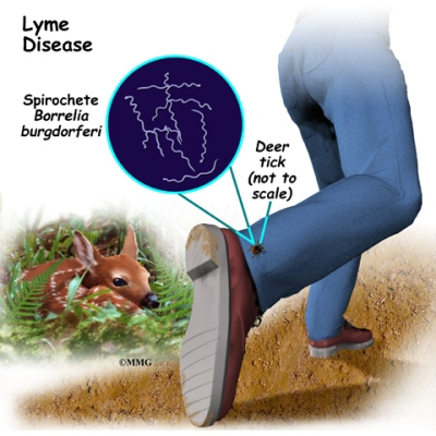
Lyme disease is a subacute inflammatory disorder caused by infection with Borrelia burgdorferi, a nonpyogenic spirochete transmitted by Ixodes scapularis (see image the end of this article), the deer tick bite, in the eastern U.S. and I. pacificus, the western black-legged tick, in the western U.S. Ticks live in wooded area and survive by attaching to a host. And tick nymphs are thought to be responsible for about 90% of transmission to human beings.

Typical symptoms include fever, headache, fatigue, and a characteristic skin rash called erythema migrans. Variations in clinical features or severity from one patient to another may be due to inborn variations in immune response, perhaps linked to the human lymphocytic antigen system. The specific symptoms are depend on stages.

First Stage : Early localized infection

  • Symptoms occur several days to month following the bite.
  • Flulike symptoms
  • Small red pimple that becomes ring-shaped rash (see image on the end of this article), and it can be large or small
Second Stage: Early disseminated infection
  • Symptoms occurs several week following the bite.
  • Neurological complication
  • Joint pain
  • Cardiac complication
Third Stage: Late persistent infection
  • Large joints becomes involved
  • Arthritis progresses

CARE INTERVENTIONS:
  1. Remove the tick with tweezers gently, wash skin with antiseptic.
  2. Take a blood test for 4 – 6 weeks following a bite to detect the presence of lyme disease.
  3. Antibiotics as prescribed if the lyme disease is confirmed.
  4. Avoid area that contain ticks (wood area, grassy area) especially in summer.
  5. Wear long-sleeved tops long pants, close shoes and hat while outside
  6. It is recommended to spray the body with tick repellent before going outside.

Images are here :


ring-shaped rash


Ixodes scapularis, the primary vector of Lyme disease in eastern North America


Nymphal and adult deer ticks can be carriers of Lyme disease.


Read more...

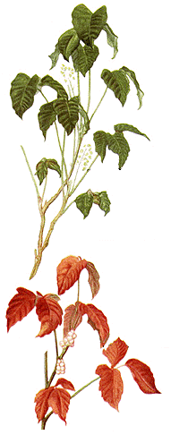
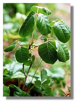
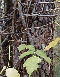
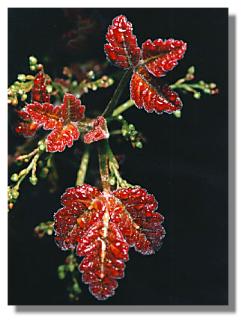
POISON IVY, OAK AND SUMAC


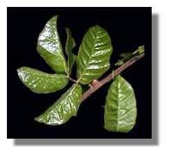
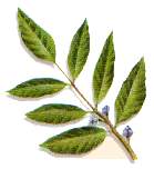
This is a dermatitis that develops from contact with uroshiol from poison ivy, oak, or sumac plant. Poison ivy, oak and sumac are most common allergy in the country claiming half the population and sensitivity to uroshiol can develop at any time. And a person who contacts with ivy, oak or sumac appears to react slightly different to all the remedies.

Assessment :

How is the nursing intervention?

When a person is suspected contact with poison ivy, oak or sumac, do these steps:
  • Cleanse the skin of the plant oils
  • Apply cool, wet dressing with Burow’s solution to relieve itching
  • Apply lotion or topical corticosteroid as prescribed, and
  • Oral corticosteroid for severe reaction as prescribed


Pictures of Ivy :


Pictures of Oak :


Pictures of Sumac :


Read more...

Contact Dermatitis


Contact dermatitis is an inflammatory response of the skin that produces skin changes after contact with a specific allergen (allergic contact dermatitis) or irritant (nonallergic contact dermatitis). It is also called a T lymphocyte-mediated dermatitis (type IV hypersensitivity).

Specific allergen that cause contact dermatitis in many people include "poisonous" plants such as poison ivy, certain foods, some metals, cleaning solutions, detergents, cosmetics, perfumes, industrial chemicals, and latex rubber.

A person who has contact dermatitis will get sign and symptom of pruritus and burning, erythema at the point of contact, edema, vesicles with drainage, as well as sign of infection.


Nursing intervention for client with diagnose of contact dermatitis will include :
  • Elevation the extremity to reduce edema
  • Maintaining a cool environment
  • Apply cool and wet dressing and tepid bath
  • Protection of the affected area from trauma
  • Prevention of scratching and rubbing of the affected area
  • Assisting with skin testing to determine allergen
  • Encourage client to avoid contact with the allergen and harsh soaps
  • Encourage client to avoid using heating pads or blankets
  • Administering (as prescribed) antibiotic, antipruritic, antihistamine, and corticosteroids.


Read more...

NCLEX - CGFNS : Skin Cancer

Skin cancer is a malignant lesion of the skin which may or may not metastasize. Skin cancer can be caused by chronic friction and irritation to the skin area and exposure to ultraviolet.

Skin cancer can be diagnosed by a skin biopsy that is positive for cancer cell.

There is three types of skin cancer: basal cell, squamous cell, and malignant melanoma. Basal cell is the most common type, arises from the basal cell contained in the epidermis. Squamous cell cancer is the second most common of skin cancer that is a tumor of the epidermal keratinocytes and can infiltrate surrounding structure, metastasize to lymph nodes and bi subsequently fatal. Malignant melanoma is a cancer of the malanocytes that can metastasize to the brain, lungs, bones, liver and skin. Malignant Melanoma is ultimately fatal.

Skin cancer can be assessed by :

  • Pruritus
  • Local soreness
  • Change in color, size, or shape of lesion
  • Waxy nodule
  • An Irregular, circular, bordered lesion
  • Small, red, nodular lesion
  • An oozing, bleeding, crusting lesion.
As a professional nurse, you can instruct client with skin cancer to :
  1. Do the preventive measures
  2. Monitor for lesion that do not heal
  3. Avoid contact with chemical irritants
  4. Wear layered clothing and use sunscreening lotions with an appropriate skin protection when outdoors
  5. Avoid sun exposure between 11 AM and 3 PM
  6. Assist with surgical excision of the lesion as prescribed


Read more...

NCLEX-CGFNS : Medication Administration

Things to be remembered that Medications are given according to the "five rights"
1. Right Patient
2. Right Drug
3. Right Dose
4. Right Time, and
5. Right Route

Some references add to that a "Right Documentation" so it becomes "sixth right".

When you give medication to the client / patient, you should also check at least three times according to the Five Rights of Medication Administration.

  • When the medication container is removed from storage
  • When the medication dose is removed from the container / ampule
  • When the medication is returned back to storage

There are commons routes of Medication Administration :
  1. PO : by mouth, the medication is given orally
  2. Sublingual : the medication is given under the tongue
  3. Subcutaneous : the medication is given into adipose tissue. Usually given at a 45-degree angle with a 1.2 to 1-inch long needle.
  4. Intramuscular : the medication is given into the muscle, given at a 90-degree angle with a long needle to pass through adipose tissue and enter the muscle.
  5. Intravenous : the medication is given into vein
  6. Topical : the medication is given on the skin
  7. Rectal : the medication is given into the rectum
  8. Intrathecal : the medication is given into the spinal canal.


Read more...