Cardiogenic Shock

Cardiogenic shock is failure of the heart to pump adequately results in reducing cardiac output and compromising tissue perfusion. The treatment is focused to maintain tissue oxygenation and perfusion and improve the pumping ability of the heart.

Signs and Symptoms:

  • Hypotension
  • Urine output less than 30 mL/hour
  • Poor peripheral pulses
  • Cold, clammy skin
  • Pulmonary congestion
  • Tachycardia
  • Chest discomfort
  • Disorientation, restlessness, and confusion


Nursing Intervention:
  • Administer morphine sulfate intravenously as prescribed to decrease pulmonary congestion and relieve pain
  • Administer oxygen as prescribed
  • Intubation and mechanical ventilation if needed
  • Administer diuretic and nitrates as prescribed
  • Prepare for insertion of intraaortic balloon pump, PTCA or coronary artery bypass graft if prescribed
  • Monitor arterial blood gas levels
  • Monitor urinary output
  • Monitor distal pulses


Read more...

Heart Failure (part 2)

Emergency Interventions for Heart Failure:

  • Place the patient in high fowler position with the legs in a dependent position to reduce pulmonary congestion and relieve edema
  • Give oxygen in high concentration by mask or cannula
  • Intubation and ventilator support if needed
  • Suction fluids as needed
  • Assess and monitor level of consciousness
  • Monitor for vital signs closely
  • Monitor for hypotension
  • Monitor for dysrhythmias (using cardiac monitor)
  • Assess for edema in dependent legs
  • Insert foley catheter if needed
  • Monitor intake and output
  • Administer morphine sulfate as prescribed
  • Administer diuretics as prescribed
  • Administer digitalis as prescribed
  • Administer bronchodilator as prescribed
  • Administer additional inotropic medications as prescribed
  • Administer vasodilator as prescribed
  • Monitor weight
  • Assess for hepatomegaly and ascites
  • Monitor peripheral pulses
  • Analyze arterial blood gas
  • Monitor potassium level closely


Interventions Following Acute Episode of Heart Failure:
  • Assist the patient to identify precipitating risk factors of heart failure
  • Encourage patient to verbalize feeling
  • Instruct patient in the prescribed medications and notify physician if side effect occurs
  • Advice patient to avoid over-the-counter medication
  • Avoid large amount of caffeine
  • Diet: low sodium, low fat, and low cholesterol diet
  • Instruct patient regarding fluid restriction as prescribed
  • Avoid isometric activities that increases pressure in the heart
  • Provide patient with a list of potassium rich foods because diuretics can cause hypokalemia
  • Instruct patient to monitor daily weight and report signs of fluid retention such as edema or weight gain


Read more...

Heart Failure (part 1)

Heart failure is defined as an in ability of the heart to maintain adequate circulation because of an impaired pumping capability. The cardiac output is diminished and peripheral tissue is not perfused adequately. The congestion of the lungs and periphery will occur.

There are two kind of heart failure: acute heart failure that occurs suddenly, and chronic heart failure that develops over the time.

Type of Heart Failure:

  1. Right Ventricular and Left Ventricular Failure: Most of them begin with left ventricular failure and then progresses to both of ventricles. Left ventricular failure can progress to acute pulmonary edema that death will occur if not treated immediately.
  2. Forward and Backward Heart Failure: In forward failure, the output of the affected ventricle is inadequate and causes decreased perfusion to vital organs. In backward failure, blood backs up behind the affected failure that causes increased pressure in the atrium behind the affected ventricle.
  3. Low Output and High Output: in low output failure, the cardiac output is not enough to meet the demand. In high output failure, the heart works harder to meet the demand.
  4. Systolic Failure and Diastolic Failure: Systolic failure means the problem with contraction and the ejection of blood. Diastolic failure means the problem in relaxing of the heart and filling with blood.


Signs and Symptoms of Heart Failure


Right Ventricular Failure:

  • Pitting, dependent edema in the feet, legs, sacrum, back, and buttocks
  • Ascites form portal hypertension
  • Distended neck veins
  • Tenderness of right upper quadrant
  • Abdominal pain
  • Nausea and anorexia
  • Weight gain
  • Fatigue
  • Nocturnal diuresis

Left Ventricular Failure:
  • Cough with frothy sputum
  • Dyspnea on exertion
  • Paroxysmal nocturnal dyspnea
  • Orthopnea
  • Crackles on auscultation
  • Tachycardia
  • Pallor
  • Cyanosis
  • Fatigue
  • Confusion and disorientation
  • Signs of cerebral anorexia

Acute Pulmonary Edema:
  • Severe dyspnea and orthopnea
  • Tachycardia
  • Pallor
  • Bubbling respirations
  • Expectoration of large amount of blood tinged
  • Profuse sweating
  • Cold and clammy skin
  • Nasal flaring
  • Cyanosis
  • Use of accessory breathing muscle
  • Tahycpnea
  • Hypocapnia
  • Anxiety, apprehension and restlessness
Continued to Heart Failure (part-2)


Read more...

Myocardial Infarction

Myocardial infarction is a condition in which myocardial tissue is abruptly and severely deprived of oxygen. It can lead to necrosis of myocardial tissue if blood flow is not restored.

Physical changes in the heart do not occur until 6 hours after the infarction, the infarcted area becomes blue and swollen, and yellow and gray after 48 hours. By 8-10 days, granulation tissues form, and develop into scar over 2-3 months.


There are three location of Myocardial Infarction:
  1. Anterior or septal MI or both: due to obstruction of the left anterior descending.
  2. Posterior wall MI or lateral wall MI: due to obstruction of circumflex artery.
  3. Inferior MI: due to obstruction of the right coronary artery

Risk Factors of Myocardial Infarction:
  • Coronary artery disease
  • Atherosclerosis
  • Smoking
  • Elevated cholesterol levels
  • Hypertension
  • Obesity
  • Impaired glucose tolerance
  • Stress
  • Physical inactivity


Diagnostic Procedures for MI:

  • Total Creatine Kinase Level: rises with 3 hours and peaks within 24 hours
  • CK-MB isoenzyme: peaks within 18-24 hours after the onset of chest pain and returns to normal 48-72 hours later
  • Troponin level: rises within 3 hours and remains elevated up to 7 days
  • Myoglobin: rises within 1 hour, peaks in 4-6 hours, and returns to normal within 24-36 hours
  • LDL level: rises 24 hours after MI, peaks 48-72 hours, and normal in 7 days
  • White blood cell count: elevated WBC on 2nd day of MI
  • Electrocardiogram: ST segment elevation, T wave inversion, and abnormal Q wave


Signs and Symptoms of MI:
  • Pain: crushing substernal pain, radiate to the jaw, back and left arm. Pain occurs without any causes primarily early morning and unrelieved by rest or nitroglycerin and last 30 minutes or longer.
  • Diaphoresis
  • Nausea and vomiting
  • Dysrhythmias
  • Dyspnea
  • Feeling of fear and anxiety
  • Pallor, cyanosis, coolness of extremities


Complication of MI:
  • Heart failure
  • Dysrhythmias
  • Pulmonary edema
  • Thrombophlebitis
  • Cardiogenic shock
  • Pericarditis
  • Mitral valve insufficiency
  • Ventricular rupture
  • Postinfarction angina


Intervention in Acute Stage:
  • Assess a description of the chest pain
  • Assess vital sign
  • Assess and monitor cardiovascular status
  • Administer oxygen at 2-6 L/min by nasal cannula
  • Establish IV access
  • Administer nitroglycerin as prescribed
  • Administer morphine sulfate as prescribed (if unresponsive to nitroglycerin)
  • Obtain a 12-lead ECG
  • Administer IV nitroglycerin and antidysrhythmias as prescribed
  • Monitor thrombolytic therapy
  • Monitor for signs of bleeding
  • Administer beta blocker as prescribed
  • Monitor for cardiac dysrhythmias
  • Monitor for complication of Myocardial infarction
  • Assess and monitor distal peripheral pulses and skin temperature
  • Monitor intake and output
  • Assess respiratory rate and breath sounds
  • Monitor blood pressure closely

Intervention Following Acute Stage:
  • Bed rest for the first 24-36 hours, allow patient to stand to void or use a bedside commode
  • Provide range of motion exercises
  • Monitor the complication of MI
  • Encourage patient to verbalize feeling.


Read more...

Angina


Angina is severe chest pain resulting from myocardial ischemia. It is caused by inadequate myocardial blood and oxygen supply, obstruction of coronary blood flow, coronary artery spasm, and a condition that increases myocardial oxygen demand. There is an imbalance between oxygen supply and demand.

The treatment is focused to provide relief of an acute attack, correct the imbalance between myocardial oxygen supply and demand, and prevent the progression of the disease and further attack to reduce risk of myocardial infarction.

Types of Angina:
  1. Stable Angina: it is also called exertional angina, occurs with activities that involve exertion or emotional stress and is relieved with rest or nitroglycerin. It has stable pattern of onset, duration, severity, and relieving factors.
  2. Unstable Angina: it is also called preinfarction angina, occurs with an unpredictable degree of exertion or emotion and increases in occurrence, duration and severity over time. The pain of this angina may not be relieved with nigroglycerin.
  3. Variant Angina: it is also called Prinzmetal’s or vasospastic angina. It is results from coronary artery spasm and may occur at rest. ST segment elevation is noted on the electrocardiogram.
  4. Intractable Angina: it is a chronic and incapacitating angina that is unresponsive to interventions.
  5. Preinfarction Angina: It is associated with acute coronary insufficiency and lasts longer that 15 minutes. Preinfarction angina is a symptom of worsening cardiac ischemia.
  6. Postinfarction Angina: It occurs after an myocardial infarction, when residual ischemia may cause episodes of angina.

Sign and Symptoms of Angina:

  • Pain: slowly or quickly, mild or moderate, last lest than 4 minutes and relived by nitroglycerin or rest. Pain may radiate to the shoulders, arms, jaw, neck, and back. Pain is substernal, crushing, and squeezing.
  • Pallor
  • Dyspnea
  • Sweating
  • Dizziness and faintness
  • Hypertension
  • Palpitations and tachycardia
  • Digestive disturbances

Diagnostic Procedures for Angina:
  • Electrocardiogram: Normal during rest. ST depression or elevation and/or T wave inversion during an episode of attack.
  • Stress Test: chest pain or changes in ECG or vital signs during testing indicate ischemia
  • Cardiac Enzymes and Troponins: Normal in angina
  • Cardiac Catheterization: to provide a definitive diagnosis.

Immediate Management:
  • Assess pain
  • Provide bed rest
  • Oxygen at 3 L/min by nasal cannula
  • Administer nitroglycerin as prescribed to dilate coronary arteries and relieve pain
  • Obtain a 12-lead ECG
  • Continuous cardiac monitoring


Read more...

Coronary Artery Disease

Coronary artery disease is a condition in which one or more coronary arteries become narrowed or obstructed. It can be result from atherosclerosis and an accumulation of lipid-containing plague in arteries. This condition causes decreases perfusion of myocardial tissue and inadequate myocardial oxygen supply. The goal of treatment is to alter the atherosclerotic progression.

Coronary artery disease leads to hypertension, angina, dysrhythmias, myocardial infarct, heart failure, and death.


Coronary artery narrowing is significant if the lumen diameter of the left main artery is reduced at least 50% or if any major branch is reduced at least 75%.

Sign and Symptoms Coronary Artery Disease:
  • Chest pain
  • Palpitations
  • Dyspnea
  • Syncope
  • Excessive fatigue
  • Cough or hemoptysis
  • Possibly normal findings during asymptomatic

Diagnostic Procedure for Patient with Coronary Artery Disease:

  • Electrocardiogram: ST segment depression or T wave inversion when blood flow is reduced and ischemia occurs. ST segment elevation followed by T wave inversion when infarction occurs.
  • Cardiac Catheterization: It will show the presence of atherosclerotic lesions.
  • Blood Lipid Levels: may be elevated.

Nursing Interventions:
  • Inform patient regarding the purpose of diagnostic medical and surgical procedures
  • Assist and identify risk factors that can be modified
  • Instruct the patient regarding a low-calorie, low sodium, los cholesterol, and low fat diet with an increase in dietary fiber
  • Inform the patient that dietary changes are not temporary but maintained for life
  • Instruct the patient regarding prescribed medications
  • Instruct the patient regarding exercise, smoking reduction, and stress reduction

Surgical Procedures for Coronary Artery Disease:
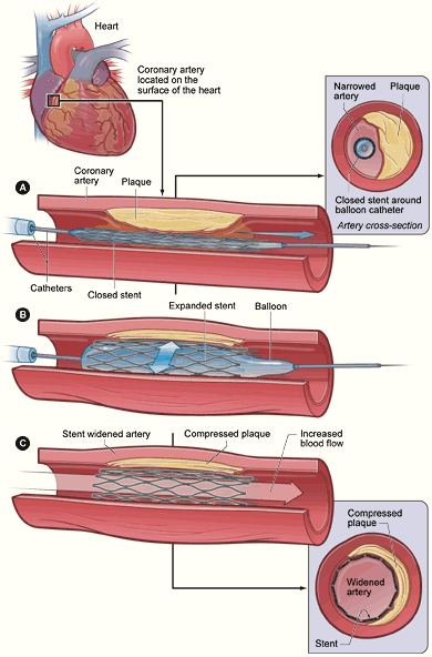
  • PTCA: to compress the plaque against the wall of the artery and dilate the vessel
  • Laser angioplasty: to vaporize the plaque
  • Vascular stent: to prevent the artery from closing and to prevent restenosis
  • Atherectomy: to remove the plaque from the artery
  • Coronary artery bypass graft: to improve blood flow to the myocardial tissue


Medications
  • Nitrates: to dilate the coronary arteries and to decrease preload and afterload
  • Cholesterol-lowering medications: to reduce the development of atherosclerotic plaques
  • Calcium channel blockers: to dilate coronary arteries and reduce vasospasm
  • Beta blocker: to reduce blood pressure


Read more...